Anatomy Phantoms
Body Part X-Ray Phantoms
Our body part X-ray phantoms allow repeated X-ray imaging of specific body regions. These phantoms include real human bones. They are ideal for schools and education, but also for medical applications or manufacturer equipment testing.
The bones are embedded in tissue equivalent material. Phantoms could be coated with opaque colour to hide the inner structures. All phantoms are hand made and unique. They may differ in size and shape. Due to production technology, there may be discolouration and cracks inside the phantom. This is related to production and represents no lack of quality. Anatomy phantoms are only sold against a proof of medical use.
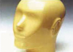
Dental Anatomy Head Phantom
Art. No. 12701 (transparent)
Art. No. 12701-o (opaque)
 The dental anatomy head is specifically prepared to be used for dental applications
such as panoramic, cephalometric or dental Cone-Beam CT/D3. Its features include:
The dental anatomy head is specifically prepared to be used for dental applications
such as panoramic, cephalometric or dental Cone-Beam CT/D3. Its features include:
- Skull with lower jaw and 5 cervical vertebrae.
- Jaw slightly open.
- Standard version: opaque (optional in transparent).
- Features 1 tooth repair and 1 inlay (or as per customer requirement).
- M6 screw hole for optional tripod positioning.
- Case for transport and storage.
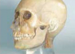
Anatomy Head Phantom I
 Art. No. 12702 (transparent)
Art. No. 12702-o (opaque)
- Skull with lower jaw and 5 cervical vertebrae.
- Connecting jaws.
- Standard version: opaque (optional in transparent).
- Case for transport and storage.
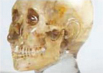
Anatomy Head Phantom II
 Art. No. 12703 (transparent)
Art. No. 12703-o (opaque)
- Skull with lower jaw, no extra vertebrae.
- Connecting jaws.
- Standard version: opaque (optional in transparent).
- Case for transport and storage
Hand Phantom
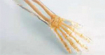 Features wrist joint (transparent / opaque)
Foot Phantom
Features ankle joint (transparent / opaque)
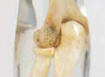
Arm Phantom
Features lower arm and elbow (transparent / opaque)
 Knee Phantom
Features thigh, lower leg and kneecap (transparent / opaque)
Spine Phantom
Features 24 vertebrae and sacral bone (transparent / opaque)
Hip Phantom
Features pelvis, 2 lumbar vertebrae and thigh part (transparent / opaque)
CT Torso Phantom
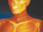 A one-piece anthropomorphic torso phantom with anatomical structures allows various CT approaches
including helical scanning. Along with state-of-the-art synthetic bones, brain with cerebral
ventricles, eye balls, lung with 3-dimensional pulmonary vessels, trachea, liver with portal and
hepatic veins, kidneys, gallbladder, pancreas, splean, aorta, cava, ureter, urinary bladder, prostate,
rectum, and sigmoid colon are embedded. Each individual organ has a particular Housfield unit,
which corresponds to the human equivalent. The original phantom material, with radiation absorption
approximate to human tissues, allows scanning in actual clinical settings.
A one-piece anthropomorphic torso phantom with anatomical structures allows various CT approaches
including helical scanning. Along with state-of-the-art synthetic bones, brain with cerebral
ventricles, eye balls, lung with 3-dimensional pulmonary vessels, trachea, liver with portal and
hepatic veins, kidneys, gallbladder, pancreas, splean, aorta, cava, ureter, urinary bladder, prostate,
rectum, and sigmoid colon are embedded. Each individual organ has a particular Housfield unit,
which corresponds to the human equivalent. The original phantom material, with radiation absorption
approximate to human tissues, allows scanning in actual clinical settings.
Full Body X-Ray Phantom
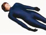 This model is unique in the world and provides best opportunities for X-ray training.
It is a must-have for all radiological schools. The phantom can be used for positioning practice
as well as for general X-ray training. The model contains a real human skeleton and allows
taking real X-ray images comparable to a real patient. In addition to the real skeleton, the
phantom incorporates reduction of heart, lungs, larynx and kidneys appearing in the X-ray images.
Each model is hand-made and differs in size and design. Phantoms may include pathologies and may
also differ in appearance. Life size.
This model is unique in the world and provides best opportunities for X-ray training.
It is a must-have for all radiological schools. The phantom can be used for positioning practice
as well as for general X-ray training. The model contains a real human skeleton and allows
taking real X-ray images comparable to a real patient. In addition to the real skeleton, the
phantom incorporates reduction of heart, lungs, larynx and kidneys appearing in the X-ray images.
Each model is hand-made and differs in size and design. Phantoms may include pathologies and may
also differ in appearance. Life size.
Lung Cancer Screening Phantom
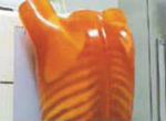 This phantom is an adapted CT phantom developed to optimise radiation dose and other scanning conditions
for Lung Cancer Screening CT examinations. Helical CT or MDCT can be tested. The phantom is designed
to simulate conditions for early detection of small lung cancers such as GGA. Quantitative evaluation on
radiation dose and density curve of the image can be done simultaneously with a single scan. The model
consists of a life-size torso with arms-up position and has the following internal structures:
This phantom is an adapted CT phantom developed to optimise radiation dose and other scanning conditions
for Lung Cancer Screening CT examinations. Helical CT or MDCT can be tested. The phantom is designed
to simulate conditions for early detection of small lung cancers such as GGA. Quantitative evaluation on
radiation dose and density curve of the image can be done simultaneously with a single scan. The model
consists of a life-size torso with arms-up position and has the following internal structures:
- bones.
- simulated tumors on sections of three lung areas (apical portion, bifurcation of the trachea, base of lungs.)
- central dose meter inserts.
- 8-step linearity phantom (8 steps, 30mm diameter).
- embedded density samples.